


Gamma Spectroscopy

Gamma spectroscopy is a technique used to measure the energy and intensity of gamma rays emitted from a sample. This technique is often used to measure nuclear radiation. In fact, gamma rays act as a fingerprint for radioactive materials and radiation interactions.
As a gamma ray moves through a detector, it will instigate a series of reaction events leading to the production of many subsequent charged particles (mostly electrons with some positrons) and secondary photons (a combination of gamma rays, X-ray, visible photons, etc.). While it is hard to quantify individual events, these events combine to produce a complicated spectrum.
Gamma spectroscopy systems need multiple components to convert incoming gamma radiation into a useful energy spectrum. This includes a suitable detector, a pre-amplifier and electronics to shape, digitize and sort (or "bin") each signal. Older gamma spectroscopy systems use a physical amplifier followed by a multichannel analyzer (MCA). However, newer systems include the functionality of the amplifier within the MCA for full digital pulse processing.
Each pulse is then added to a histogram depending on the energy level of the gamma ray, which over time builds to an energy spectrum.
How Does Gamma Spectroscopy Work?
Turning a series of gamma rays into a readable spectrum takes a chain of hardware, with each stage handling a specific job. The components needed for this include a voltage supply, detector, a pre-amplifier, then various processing and shaping electronics. These electronics can be built around either an analog or digital signal-processing chain.
High Voltage Supply
Many detectors need a high voltage supply. In semiconductor detectors, the voltage supply provides an electric field to guide the electron-hole pairs out of the detector and into the pre-amplifier.
Detector
Detectors are where the signals are absorbed and collected. A signal from the source or sample comes straight into the detector where ideally it is absorbed.
There are two main types of gamma ray detector: scintillators and semiconductors. In scintillators, gamma radiation produces light signals that are then collected and converted into an electrical signal using photomultiplier tubes or other photodetector systems. In semiconductor devices, photons are absorbed and converted into free charge within the semiconductor material. To collect these, an applied voltage drives these charge carriers to opposing poles.
Semiconductor Detectors
Semiconductor (SC) materials or solid-state detectors, such as high-purity germanium (HPGe), are a popular choice for radiation detection systems as they have a much higher mass density and atomic number than their scintillator counterparts. This means that semiconductor detectors can be smaller while maintaining high detection efficiency. They can also achieve higher energy resolution than scintillators, generating more information per unit of deposited energy. Also, they have a fast response time, and it is easy to modify their thickness/geometries depending on application. However, they can be vulnerable to radiation damage and their maximum area is limited. They must also be cooled to low temperatures to supress thermal noise and maximize efficiency.
In an SC detector, an incoming photon will create many electron-hole pairs as it travels through. The number of electron-hole pairs depends on the ionization energy of material which is usually quite low for SCs (~3 eV). The applied bias across the depletion region drives created charge carriers to the edge of the active area. These can then be collected and analyzed.
Scintillation Detectors
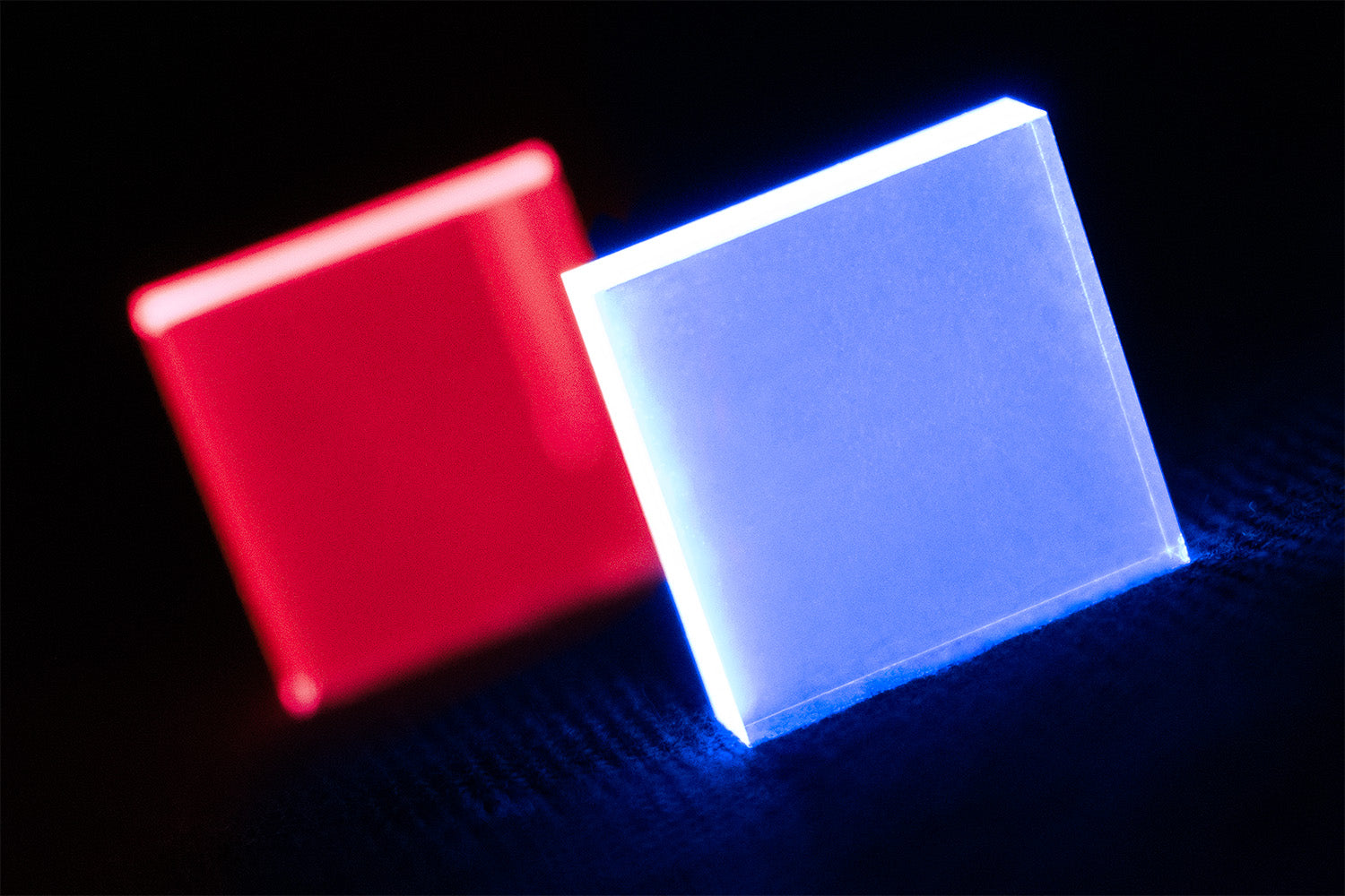
The other type of detector is a scintillator. In scintillation detectors, radiation is absorbed by the scintillator material and reemitted as a visible light signal. These signals travel through the material, towards a photodetector or photomultiplier system. This multiplies the signal, turning it into a significant electrical output.
There are two broad classes of scintillator, and they generate light through different physical mechanisms.
For inorganic scintillators (such as doped alkali halide crystals like NaI(Tl) or CsI(Tl)), their intrinsic bandgap usually falls outside the visible range, but introducing a small amount of dopant creates additional energy levels, known as luminescence or recombination centers. Free electrons can migrate to these dopant sites, where they relax emitting a photon. Inorganic scintillators typically achieve much higher light output than their organic counterparts, but have a slower response time, generally tens to hundreds of nanoseconds rather than a few ns.
Organic scintillators include crystals like anthracene and stilbene, as well as plastic and liquid scintillators. They are cost-effective and can create large-area detectors. For example, in plastic scintillators, a small amount of organic scintillator material is added into a PMMA or PVT matrix. This polymerized matrix can be any size and be grown into a range of different architectures.
Scintillator materials also have a shorter decay time than semiconductors and are less prone to radiation damage. However, converting incoming radiation into an electrical signal using an organic scintillator relies on several low-yield steps. Their light yield tends to be relatively low, and they generally aren't efficient at detecting high-energy or heavier particles. However, inorganic scintillators are widely used in gamma spectroscopy.
Detector Design
The ideal detector material and system design depends largely on the desired application. Detectors that prioritize fast signal collection are good for radiation counters, and those with high energy resolution are good for radiation spectroscopy.
The measurement ranges of the detector are determined by multiple factors. Its energy capability is limited either by the absorption co-efficient of the detector material and the detector thickness. High energy photons are likely to pass right through the detector. However, with larger detectors there is a higher chance of absorption.
The lower limit of the energy capability is set by the noise floor or by the lower energy X-ray being absorbed in the entrance window and dead layer before reaching the active volume.
The measured signal is also affected by where the photon absorption takes place within the detector. This impacts the signal, the shape of the detector pulse and the charge collection time, which in turn influences the processing electronics needed and affects counting rates. Fast count rate is not the most important factor for gamma spectroscopy, so most systems prioritize energy resolution.
The detector is connected to a pre-amplifier, which collects the charge and converts it into a voltage for the electronics to process.
Shielding
Shielding the detector is very important. This minimizes the influence of ambient radiation. This could be lead, steel, cadmium or copper. This material needs to be non-radioactive and highly absorbing. For low level systems, 15 cm lead is suitable.
However, X-ray fluorescence can be generated from the lead shielding, so the inside should be lined with successive layers of decreasing atomic number, typically tin followed by copper.
Electronics
All spectroscopy systems require certain components: a pre-amplifier, pulse shaping systems, an analog-to-digital converter and some sort of multichannel analyzer.
The electronics involved with gamma spectroscopy have taken many forms. Originally, these were large inbuilt systems where all of the pulse shaping and processing took place within a large unit.
Then, there was a move to more modular systems where each physical element is connected in a series of slots to build a spectroscopy system. These elements are designed to fit into a standard Nuclear Instrumentation Module (NIM) crate. This makes it easier to replace these physical elements when they succumb to drift due to wear. This also means that each feature can be interchanged or adapted to suit the experiment at hand.

Now many systems are being introduced with digital pulse processing. This negates the need to rely on solely physical components for things like pulse shaping, baseline correction and pile-up rejection. Physical amplifiers can drift and misalign with age and varying temperature, creating inconsistencies between measurements. Furthermore, digital systems can introduce more sophisticated filtering techniques, and dual pole-zero cancellation functions, improving resolution, stability, and throughput.
Pre-Amplifier
This is the element that goes between the detector and the amplifier or digital multichannel analyzer. Despite its name, its main role is not to amplify a pulse, but to collect charge and send it to the processing system. Ideally, the height of the output pulse should be proportional to the energy of the absorbed gamma ray.

The pre-amplifier must act as a high-impedance load for the detector and a low-impedance source for the amplifier, facilitating the flow of electricity from detector to amplifier. There are charge-sensitive and voltage-sensitive elements, with the former being most commonly used in HPGe gamma spectroscopy systems.
There are two main types of charge-sensitive pre-amplifiers: feedback and reset. In feedback pre-amplifiers, a pulse excites the system, and a feedback loop slowly returns to the baseline, producing an exponential tail. However, reset pre-amplifiers allow signals to accumulate in step functions over a given time, then automatically restore to zero at given intervals (such as once the system reaches a voltage limit).
These elements can suffer from count rate overload. Especially in feedback pre-amplifiers, the exponential tails can mean that successive signals build up over time. If these signals become larger than the dynamic range of the pre-amplifier, it will saturate.
Amplifier
Analog systems need an amplifier to shape the pulse before it enters the multi-channel analyzer for analogue-to-digital conversion and plotting. In digital pulse processing systems, these functions are handled digitally within the multichannel analyzer after the signal has been converted.

The fast-rising, long-tailed output of a feedback pre-amplifier cannot directly be used to measure peak height. For example, it is difficult to differentiate two similar signals occurring in quick succession due to the long tail. This signal ideally needs to be manipulated into a Gaussian shape before the height is measured.
Amplifiers handle many functions including pile-up rejection, pole-zero cancellation, baseline restoration and, most importantly, pulse shaping.
-
RC Shaping
Amplifiers use a combination of resistors and capacitors in series-shunt combinations to shape the pulse. A differentiator or high-pass filter (CR) only allows high frequency signals through. Then the pulse is passed through integrators or low-pass filters (RC) which only allows low-frequency components of the pulse through. Combining these two filters together takes a signal from the pre-amplifier and produces a shorter pulse with a shape that can be more easily processed by ADCs.
Running the signal through several RC shaping circuits will produce a semi-Gaussian shaped output or a unipolar output.

-
Pole-Zero Cancellation
The differentiator-integrator amplifiers assume that the pre-amplifier output is basically a step voltage. This is acceptable for the most part but leads to one main error. The feedback circuit of the pre-amplifier creates a dip in the final uncorrected output. If followed closely by a second signal, the MCA could incorrectly measure the amplitude of this second signal. This can be corrected through pole-zero cancellation.
In most analogue systems, this cancellation is adjusted by eye, using an oscilloscope, and needs to be checked whenever the shaping parameters are changed.

-
Baseline Restorer
Traditional amplifiers are AC-coupled, so they cannot pass DC. Therefore, the net area of the pulse above and below baseline is equal forced to zero, which means every pulse carries an undershoot. For long tailed pulses, this is only a slight shift, but it can accumulate with multiple pulses. As the MCA measures height relative to the baseline, it is important that this baseline is as accurate as possible. These spectroscopy systems process random signals from many incidents, so the baseline is always shifting, which can limit resolution.
-
Pile-up Rejection
When two or more signals occurring almost simultaneously, they can be incorrectly summed as one larger pulse. A fast-shaping amplifier can be used to trigger an inspection interval. This element cannot measure height or any pulse detail but simply signals that a pulse has arrived. If another pulse is detected by the fast-shaping amplifier within this investigation time, the previous signal is cancelled, and neither signal is processed.
Multichannel Analyzer
A multichannel analyzer is the component that digitizes the height of each incoming pulse, processes and bins them to create a histogram. With enough signals this becomes the energy spectrum. In digital processing units, the MCA also encompasses a lot of the functionality of the amplifier, acting as an amplifier, an ADC and analyzer all in one.
The MCA receives an incoming signal from the amplifier (in analogue systems) or straight from the pre-amplifier (digital MCAs). To convert the incoming signal into a readable spectrum requires several steps including an analog-to-digital converter and an onboard processor with a digital signal processing system.
- Analog-to-digital converters (ADCs) are like the central nervous system of the multichannel analyzer. They efficiently translate pulsed signals from a radiation detector into a form that a computer processor can understand and analyze. A modern digital multichannel analyzer converts a voltage pulse waveform into a stream of digital bits using a high-speed, high-resolution ADC.
- After this, the processor determines the signal height and sorts all the detected pulse amplitudes over time in their respective bins. Each individual pulse is added to the memory location of the channel matching the pulse height. This provides users with a spectral picture of the radiation source.
Analog MCAs contain the following components:
- Linear gate or single channel analyzer, that only lets signals within a certain pulse height through, rejecting all pulses outside that window.
- Input gate, which only lets one signal at a time through.
- ADC
- Memory count
Digital MCAs use an ADC straight after the pre-amplifier to immediately convert the signal into a digital pulse. This signal can be shaped, processed and binned mathematically. Algorithms are used to perform fast trigger, baseline restoration, pile-up rejection, trapezoid shaping and pole-zero cancellation. This creates a trapezoidal signal where the height represents gamma ray energy. This signal can be easily processed by the histogram memory. The trapezoidal signal is easier for the system to read as it has an extended flat maximum at the pulse height, rather than the top of a near Gaussian distribution. Also with digital manipulation, processes like pole-zero cancellation can be mathematically calculated to compensate for multiple pole corrections. This leads to more accurate pole-zero corrections than can be achieved by eye.
Analyzing Gamma Spectra
It can be difficult to analyze gamma spectroscopy spectra as a single interaction creates a cascade of secondary events that happen in a short period of time. However, there are some characteristic features that you can identify in a gamma ray spectrum.
The photoelectric effect dominates below a few hundred keV and pair production dominates above several MeV, with Compton scattering dominant in between.
None of these interactions are independent. Therefore, the full spectrum will represent a mix of many of these events. For this reason, analysis requires careful consideration.
Photopeaks
In the photoelectric effect, one of the inner electrons in a detector atom absorbs a gamma ray. Once this electron is ejected from the atom, another outer orbit electron relaxes to fill this hole, releasing a photon.

This X-ray is usually reabsorbed within the detector, so the whole energy of the incident gamma ray is deposited in the active volume. Occasionally the X-ray escapes instead.
Complete deposition produces a photopeak at an energy equal to the full energy of the incident gamma ray. The photoelectron itself carries away the gamma ray energy minus the shell binding energy, and the X-ray or Auger cascade that follows deposits the remainder nearby. If the characteristic X-ray escapes, the event instead lands in an X-ray escape peak, at an energy equal to the photopeak energy minus the X-ray energy.
These photopeaks are the sharp peaks that you can see in most gamma spectra. This will provide an identifiable “finger print” of that material. In order for these peaks to be visible in the gamma spectrum, the gamma or X-ray must be completely absorbed by the detector.
Identifying these photopeaks is the key to gamma spectroscopy. Different radioactive materials will have various signatures that you can identify in a spectrum. Standard gamma emission peaks for most elements are well documented in several locations.
However, there will also be other peaks from unknown sources. Some causes for non-standard peaks include the summation of gamma rays emitted simultaneously (such as cascade peaks) or emissions from naturally occurring radioactive materials (NORM).
Naturally Occurring Radioactive Materials
There are radioactive materials all around us, including in gamma spectroscopy systems. Although most of them have quite long half-lives, the sheer number of these atoms means these decays happen relatively often. These come from cosmic-ray interactions in the atmosphere, cosmic-ray muons reaching the detector, as well as naturally occurring terrestrial radionuclides including Potassium-40 and the decay chains of Uranium-238 and Thorium-232. Also, radon gas is itself radioactive and produces a chain of short-lived radioactive progeny (Pb-214 and Bi-214) whose gamma emissions are often the largest contributors to background peaks in a real-world gamma spectrum. Together, these make up much of the external background radiation which will often be picked up by gamma spectroscopy.
Identifying, quantifying and removing these signals takes seriously consideration. In fact, in high-energy physics study removing as much of the background as possible is critical.
Sum Peaks and True Coincidence Summing
When an intermediate nuclear energy state in a decay chain has a short lifetime, two decay processes can happen in very short succession. This may be interpreted as one gamma emission with higher energy, but this energy will be equal to the summation of the two decay emissions.
For example, when 60Co decays into 60Ni , it must go through a beta decay step, then two gamma decay steps. The two gamma decays have energies of 1.173 MeV and 1.332 MeV, but as they happen in quick succession, they are sometimes counted as one emission at 2.505 MeV.
The Compton Continuum
An incoming gamma ray can scatter off a loosely bound electron in the detector, transferring part of the photon's energy to the electron as kinetic energy. The gamma ray loses some energy and is scattered in a different direction. This lost energy will be transferred to the electron, which recoils.

The energy of the resulting gamma ray changes with its deflection angle according to this equation:

This creates several recognizable features in the spectra. The sharp photopeaks will be superimposed on a continuum, created by Compton scattering. The continuum represents the varying angles and energies the scattered gamma ray will have.
This continuum drops off at 180°, where the maximum amount of energy has been transferred to an electron. This creates the characteristic Compton Continuum with a drop off at a certain point (Compton Edge).

Due to the varied nature of this feature, there isn’t much usable information to be extracted from this continuum, but it still plays with the counting statistics in the interesting region of a photopeak. This should be quantified in order to accurate count the photopeaks.
Pair Production

A high energy gamma ray (>1.022 MeV) can create an electron-positron pair near the nucleus of an atom. This positron quickly annihilates with an electron, producing two back-to-back 511eV gamma rays of equal energy. These gamma rays can be absorbed or involved in other interaction mechanisms (e.g. Compton scattering/photoelectric effect). But if they do escape, they can create a single escape or double escape peak.
With high energy radiation, pair production becomes much more prevalent. This event will appear as single or double escape peaks at:
equation
Detector Artifacts and Limitations
In the spectrum, the detector itself can also contribute artifacts to the gamma spectroscopy output.
The noise contributions of the pre-amplifier in particular can limit the resolution of the detector and lead to peak broadening. Depending on the sensitivity of your spectroscopy system, the pre-amplifier will have to detect signals as low as tens of electrons. However, in most gamma spectroscopy systems, an equivalent noise charge (ENC) measurement of 100-1000s electrons is standard.
This equivalent noise charge is also known as the front-end amplifier noise floor. Developing a pre-amplifier with low noise levels is vital for creating very sensitive gamma spectroscopy equipment.
Another contribution comes from tailing effects. After processing, some peaks may be left with an asymmetrical distribution. Incomplete charge collection due to defects in the detector can appear as tailing off at lower energies. High energy tailing can also occur if the baseline does not restore to zero before the second pulse is analyzed (i.e. at high count rates).
Limitations of Gamma Spectroscopy
-
Dead Time
Dead time represents the amount of time it takes to process a signal. From the signal entering the detector to a count being added to the histogram, the signal must be amplified, shaped, digitized and binned. This limits how quickly another signal can be allowed through to the MCA from the detector, ultimately limiting count rate of the spectroscopy system.
-
Saturation
In contrast to dead time, saturation refers to when the amount of charge in a single pulse, or as a result of the accumulation of loads of pulses in quick succession, exceeds the ADC's dynamic range. This means that each pulse looks "clipped" on the top since the voltage measured at the ADC is limited.
-
Non-Linearity
There should be a linear relationship between pulse height and channel number. This line is defined by its gradient and intercept, and is used to calibrate the spectrometer. Two related but distinct measures describe how well real hardware achieves this: integral and differential linearity. Integral linearity defines how far the channel boundaries moved from a straight line distribution, while differential linearity measured how even the channel widths are. Non-linearity is mainly a feature of the ADC, but the pre-amplifier and signal shaping chain also contribute.
Gamma Spectroscopy Applications
Broadly, gamma spectroscopy has many applications including:
- Detection of nuclear materials
- Dealing with nuclear waste
- Environmental monitoring
- Nuclear safeguarding
- Medical scans and biomolecular tracking
- Nuclear and particle physics research.
More Resources
 Scintillator Materials
A scintillator is a material that will absorb energy from incoming radiation and convert it to a visible photon emission. These are used in scintillation detectors to measure and identify specific types of radiation.
Read more...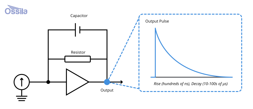 Pole-Zero Cancellation
Pole-zero cancellation is an important part of radiation spectroscopy analysis. Converting charges created from radiation events, such as gamma rays produced in gamma spectroscopy, into binnable signals introduces a shaping error which can impact later analysis.
Read more...Contributors
Written by
Product Developer
Product Developer
Diagrams by
Graphic Designer
References
- Live Chart of Nuclides, IAEA – Nuclear Data Section
- Improved sensitivity in the search for rare decays..., B. Lehnert et al., Euro. Phys. Jour. C (2025)
- Low-Noise Front-End Electronics for Semiconductor X-Ray and Gamma-Ray..., Arto Niemela, National Technical Reports Library (1998)
- The impact of high-energy tailing in high-purity germanium..., S.M. Collins et al., Applied Radiation and Isotopes (2020)
- Radiation Detection and Measurement, G.F. Knoll, John Wiley & Sons, Inc.