


Colony Counting: Method, Tips & Manual vs Automated

The Method | Best Practices | Working Principles | Manual vs Automatic Systems | Applications
Colony counting is a key technique in microbiology, offering critical insights into microbial populations. The process allows researchers to estimate the number of bacterial, fungal, or other microorganism colonies growing on solid culture plates.
As each colony originates from a single viable microorganism, colony counting is a valuable method for assessing growth patterns, antibiotic resistance, sensitivity to disinfectants, and genetic mutations. It is also used to evaluate the effectiveness of treatments like fermentation, pasteurization, sterilization, or filtration.
Despite being a straight forward method, the implications are vast, connecting raw microbial growth with actionable insights that directly influence microbiological research. While colony counting began (and still continues in many labs) as a manual technique with each colony counted by hand, there are now automated systems that speed up the process and reduce human error.
The Method
The colony counting process involves several steps:
- Sample collection and dilution: Collect samples in aseptic conditions to avoid unwanted contamination. You may need to dilute the sample to ensure colony numbers remain countable. The sample dilution formula is:
- Plating: Spread samples evenly on the agar plate using techniques such as the spread plate or pour plate method, carried out in the laminar flow hood.
- Incubation: Incubate plates under specific conditions, such as temperature, based on the target microorganisms’ growth needs.
- Counting: After incubations, count colonies using a manual or automatic colony counter.

Purpose and Importance
Colony counts directly influence the interpretation of biological phenomena used to improve experimental techniques and ensure better informed decision-making in microbiology research.

Quantification: The number of colonies allows researchers to determine the population size of the sample. This is an important step in understanding the growth characteristics and behavior of the microorganisms.
Microbial viability: The count determines the viability of the microorganisms within a sample, as only viable cells have the ability to proliferate and form colonies. This indicates their potential to either cause infections or carry out specific functions, such as fermentation.
Microbial growth: By counting colonies at different times, researchers can monitor the growth of the microorganisms. Analyzing the speed at which the microbes grow allows researchers to evaluate specific factors, such as growth-promoting and antimicrobial agents.
Quality control: Colony counting allows manufacturers to monitor contamination levels in products as well as ensuring safety standards.
Best Practices
For successful counting, the ideal number of colonies per plate is 25-300 as too many colonies affect the accuracy, while too few colonies lack statistical relevance. On top of that, when plating the sample it is important to consider the origin and the research question you want answered and set up your experiment based on these considerations.
Keep your workspace clean and safe
The first thing you should do when preparing to colony count is create (and maintain) a clean and organized workspace to reduce contamination risks, ensuring that benches, tools, and consumables remain sterile and properly arranged. To minimize human error, work methodically, clearly label tubes and plates, and double-check dilution calculations before proceeding. Alongside the whole counting process, you should always follow safety guidelines such as using appropriate PPE, disposing of biological waste correctly, and adhering to biosafety requirements.
Calibrate equipment regularly
Colony counters need to be regularly calibrated to ensure measurement accuracy and reduce systemic error. When performing calibration, it is important to follow the manufacturer's schedule along with the institutional quality-control guidelines. We recommend maintaining a calibration log to help you verify equipment performance before beginning any experiments, especially when quantitative microbiology is involved.
Follow standardized counting protocols
To ensure consistency and reproducibility, use established protocols for methods such as spread plating, pour plating, or streak plating. Adhere to defined incubation times, temperatures, and media specifications. Additionally, when counting colonies, establish appropriate rules (i.e. 30-300 colony forming units per plate for reliable quantification) that you follow throughout the experiment, and document any deviations or anomalies observed during enumeration. The use of both positive and negative controls will help validate techniques, media quality, and dilution accuracy.
Perform proper serial dilution
When performing colony counting and preparing the samples, it is important to use aseptic techniques and properly calibrated micropipettes to avoid cross-contamination or volumetric inaccuracies. To ensure the serial dilution is properly performed, mix each dilution thoroughly before transferring the aliquots to the next step. Select dilution factors that will produce countable plates and consider preparing duplicate or triplicate plates for each dilution to ensure accuracy and precision.
Document methods and results meticulously
Record all the steps of the procedure, reagent lot numbers, plate counts, dilution factors, incubation conditions, and any deviation from standard operating procedures. Use structured data sheets or electronic lab notebooks to standardize record-keeping. Include photos of plates when possible to support traceability and later review. To strengthen the confidence in the results, carry out biological and technical replicates. This also helps in identifying any outliers. It is also important to evaluate data critically by reviewing the plate counts, growth patterns, and recorded conditions for inconsistencies before finalizing results.
Working Principles
Colony counting lets researchers study the growth pattern and colony formation of microbes by converting the disordered growth into precise, measurable data. Microorganisms growing on solid agar plates are generally counted using colony-forming units (CFU). CFUs represent an estimate of viable cells that can reproduce under controlled conditions. It is based on the assumption that each viable microbe can reproduce and form colonies through binary fission under controlled conditions. The CFU per millilitre or gram is calculated based on the dilution factor and colony count.
Instruments known as colony counters assist in obtaining these vital CFU counts with a high degree of reliability and precision. Two types are currently available: manual and automatic. Manual counting of colonies can often lead to inaccuracies and can be subjective. Automated colony counting provides a reliable solution to this problem, offering a more efficient and standardized way to count colonies accurately.
Manual Counting
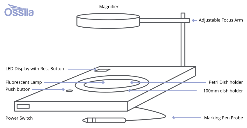
Begin by placing the petri dish on an illuminated pad designed to improve visibility. With a felt-tip pen, mark each colony directly on the dish. This triggers an electronic count, ensuring accurate tracking of the colonies. In some advanced models, magnification is included to assist in identifying small or densely packed colonies. This improves precision and reduces the likelihood of missed or overcounted colonies.
Manual colony counting often uses the following components:
- Illuminated base: The base of the counter is equipped with a bright, evenly lit surface that highlights the colonies on the petri dish.
- Pressure-sensitive surface: A pressure-sensitive pad registers and records each mark made by the researcher.
- Felt-tip pen: A specialized marking pen is used to manually highlight colonies on the petri dish. Each pen mark counts as one colony.
- Magnification lens: Some models include a built-in magnifying glass to help researchers in identifying smaller colonies.
- Digital display: More advanced manual counters may have a digital display that shows the total of counted colonies.
Advantages
Manual colony counters offer several advantages, particularly in settings that require flexibility and human expertise:
- These devices allow researchers to directly observe and mark colonies. This allows them to recognize overlapping colonies that automated counters might miss.
- As cost-effective options, they are accessible for smaller laboratories or institutions.
- Their straightforward design means that they are easy to use and maintain. They do not depend on complex software, reducing the risk of technical issues.
Limitations
While manual colony counters are useful tools, they come with several limitations:
- The process of manually marking colonies can be time consuming and labor intensive.
- They have a higher risk of human error.
- Advanced automated data analysis, such as detailed characterization of colonies by size or shape, cannot be provided.
- Manual counting is not efficient for handling large sample volumes, making it less suitable for high-throughput laboratories.
Automatic Counting
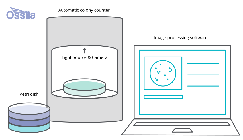
A light source illuminates the petri dish, which makes the colonies clearly visible for imaging, then a high-resolution camera captures a detailed image of the dish. This image is digitized and used as the basis for advanced computational analysis. Specialized algorithms process the image through several stages:
- Grayscale conversion simplifies the image by removing color information.
- Thresholding isolates the colonies by setting intensity levels to recognize them from the background.
- Segmentation identifies individual colonies for further analysis.
- The colonies are counted and characterized based on size, shape, and other set parameters.
Automated systems delivers results almost instantaneously, displaying them or exporting the data for further use in research or reporting.
Advantages
Automatic colony counters provide numerous advantages, particularly in enhancing efficiency, accuracy, and scalability in microbiological analysis. These include:
- Reducing the time and labor required, making them ideal for high-throughput laboratories.
- Eliminating the inconsistencies by using algorithms to accurately identify, count, and analyze colonies, even in dense or overlapping samples.
- Providing detailed data on colony characteristics, such as size, shape, and color, which can be valuable for research.
- The results are generated and displayed within seconds.
Limitations
While automatic colony counters are efficient and precise, they have limitations:
- Expensive; requiring significant investment and maintenance, which may be inaccessible to smaller labs.
- Their accuracy depends on image quality, and they can struggle with overlapping or irregular colonies that need human judgment.
- Rely on complex software, requiring trained operators and being prone to technical malfunctions.
Manual vs. Automatic Counting
Many microbiologists count colonies manually, which can be slow and time consuming. Additionally, some colonies may be too small to see clearly or may blend with color of the medium. To address these challenges, an automated colony counter offers a sustainable and efficient solution. These instruments automate the counting process, ensuring fast and more accurate results, maintaining the integrity of biological procedures.
| Automatic | Manual | |
|---|---|---|
| Method of counting | Uses digital imaging, AI/ML algorithms, or optical sensors | Human visually counts colonies |
| Accuracy | High and consistent; reduces human error | Can vary with operator skill and fatigue |
| Speed | Very fast; seconds per plate | Slow; minutes per plate depending on density |
| Reproducibility | Highly reproducible across runs and users | Low reproducibility; subjective interpretation |
| User effort | Minimal; often one-button operation | High; requires focused attention |
| Throughput | Suitable for high-volume labs and automation workflows | Suitable for low-volume or occasional counting |
| Data output | Digital records, images, metadata, exports to LIMS | Usually manual entry; limited traceability |
| Cost | High upfront cost | Low cost; little equipment needed |
| Training requirements | Some training needed for software and calibration | Basic training; simple to learn |
| Best use cases | Research labs, industrial QC, clinical diagnostics | Teaching labs, low-budget labs, simple test |
Applications
Accurate colony counting underpins experimental success in many different applications. It ensures products are safe before discharge, maintains the reproducibility of experimental results, and enables meaningful comparisons across studies.
Microbial detection: in food, water, soil, air, and pharmaceutical samples
Antibiotic susceptibility testing: to determine the effectiveness of antibiotics or severity of antibiotic resistance
Microbial ecology: studying microbial diversity and ecosystems
Research and development: exploring microbial behavior, genetic studies, and new treatments
Biotechnology: screening genetically modified organisms and testing antimicrobial agents
Learn More
 Troubleshooting Your Colony Counting
While colony counters enhance efficiency and precision, they can present challenges impacting the accuracy and workflow of the experiments. Understanding common issues and how to solve them aids in reliable results.
Read more...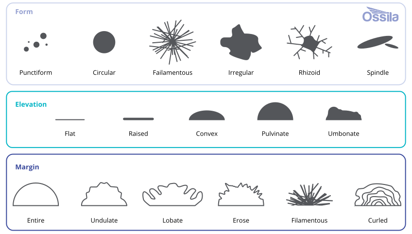 Colony Morphology
Colony morphology describes the characteristics of cultures. It refers to characteristics visible to the naked eye of both bacterial and fungal colonies growing on an agar plate. It helps researchers to choose the appropriate confirmatory tests. While the methods might look simple, careful observation and examination of colony characteristics are essential in microbiology.
Read more...References
- R. S. Khandpur Colony Counter, Automated Wiley online Library, Chapter 95, 2019
- W.B. Chen & C. ZhangAn automated bacterial colony counting and classification system Springer Nature, vol. 11, pp. 349-268, 2009
- E. Heuser, K. Becker & E.A. IdelevichEvaluation of an automated system for the counting of microbial colonies Microbiology Spectrum, vol. 11, 2023