What is Spectroscopy? Definition and Types


Spectroscopy is the study of the interaction between a radiative energy and matter and can be performed using a range of techniques.
There are several different types of spectroscopy. Generally, in spectroscopic studies, radiative energy is recorded by a spectrometer after it interacts with, or is emitted by, the material being studied. The spectrometer outputs the information as a spectrum, which shows the intensity of the radiation as a function of energy, frequency, or wavelength. These spectra are then used to obtain information on important properties of the material, including physical or chemical structure, composition, and concentration.
Different types of radiative energy used in spectroscopy include electrons, neutrons, ions, and acoustic waves. However, by far the most widely studied type is electromagnetic radiation in the UV, visible, and IR regions. For this reason, the term "spectroscopy" is often interchangeably with "optical spectroscopy".
Optical spectroscopy has applications in numerous fields of scientific research including materials science, biomedicine, astrophysics, and environmental analysis. Depending on the experimental set up, it can be used to measure absorption, transmission, reflectivity, scattering emission (photoluminescence and fluorescence), Raman scattering (via Raman spectroscopy), and more.
Spectroscopic methods can be categorized by the type of radiation that they measure (e.g. IR, UV, VIS) or by the interaction being measured (e.g. absorption, emission, scattering, fluorescence). Common types of spectroscopy include IR and NIR spectroscopy, UV-VIS spectroscopy, and Raman spectroscopy.
What is Electromagnetic Radiation?
Electromagnetic (EM) radiation consists of oscillating electric and magnetic fields that propagate through space as a wave. The fields oscillate perpendicular to one another and to the direction of propagation, as shown in the figure below. The propagating waves allow the transfer of energy at a speed of 299,792,458 meters per second (often rounded to 3x10⁸ ms-1) in a vacuum.
According to the theory of wave-particle duality, electromagnetic radiation can act both as waves, and as particles. Particles of light, known as photons, are massless and uncharged. Each photon has an energy, E, given by:
where h = 6.626x10⁻³⁴ Js (Joule seconds) is Planck's constant and v is the frequency of oscillation of the electric and magnetic fields, which are synchronous.
Photons can also be characterized by their wavelength, λ, which is the distance between successive peaks and is given by:
Different wavelengths of light correspond to different regions of the electromagnetic (EM) spectrum, with light of wavelength ~400-750 nm corresponding to the visible spectrum. Within the visible spectrum, different wavelengths are observed as different colors, with 400 nm corresponding to violet, and 750 nm to red light.
Optical spectroscopy generally concerns light in the ultraviolet (UV), visible and near-infrared (NIR) regions of the electromagnetic spectrum. The UV range of the spectrum covers from 10 nm to 400 nm, and NIR light covers from 750 nm to 1,400 nm.
The UV range can be further divided into several sub-categories. The closest to the visible region - and undoubtedly the most well-known - are UVA (315-400 nm) and UVB (280-315 nm). These are able to penetrate the Earth's ozone layer and reach its surface. UVC (100-280 nm) is almost completely absorbed by the Earth's atmosphere and only a very small amount is able to reach the Earth's surface. UV radiation with wavelengths of 10-200 nm is only able to propagate inside a vacuum and is hence known as vacuum UV or VUV.
Spectroscopy can also be performed at wavelengths outside this range; for example, in the x-ray or microwave regions.
Electron Shells and Orbitals
In atoms, electrons exist in electronic orbitals within electron shells. According to the Aufbau principle, the electrons must start from the orbital of the lowest energy and then fill the electron shells in order of increasing energy. The number of electrons in each atom will depend on the atomic number of the element involved, i.e. the further down in the periodic table an element is, the more electrons it has. For example, a carbon atom, which has an atomic number of 6, has 6 electrons.
The lowest energy state of an atom or molecule is one in which all occupied shells are full; every orbital in every occupied shell has two electrons and all higher energy shells are empty. In order to achieve the lowest energy state, therefore, atoms with partially-occupied shells will form bonds with other atoms to form compounds with fully-occupied shells.
All atoms and compounds have their own electronic structure, arising from the allowed energy levels of electrons within the material. It is this electronic structure that is studied using optical spectroscopy.
Absorption and Emission of Photons
When a material absorbs a photon, an electron is promoted from a lower to a higher energy level, e.g. from its ground state, E0, to its first excited state, E1. Conversely, when an electron relaxes from a higher to a lower energy level, a photon is emitted. In both cases, the wavelength of the photon, Eph, will be related to the energy gap between the two energy levels according to E=hc/λ (see electromagnetic radiation). These are known as electronic transitions, and the energy gap between the two levels generally corresponds to a photon in the UV or visible region of the electromagnetic spectrum.
These processes are demonstrated in the diagram above. The left-hand diagram shows the promotion of an electron from the ground state, E0, to the first excited state, E1, through the absorption of a photon with wavelength λ and energy Eph. This is equal to the energy gap between the two levels. The right-hand diagram shows the subsequent emission of a photon of the same wavelength and energy as the electron relaxes back down to the ground state.
Absorption and emission of photons can also occur when electrons transition between vibrational and rotational states. Vibrational transitions in molecules occur due to the bending and stretching of bonds, and these involve photons in the near-infrared and mid-infrared part of the electromagnetic spectrum. In this case, the transition will occur within a single electronic state. Rotational transitions involve photons in the far-infrared (lower energy) and are due to the quantised changes in the angular momentum of molecules. Rotational transitions occur within the same vibrational state (with the exception of ro-vibrational transitions).
IR Spectroscopy and NIR Spectroscopy
IR and NIR spectroscopy study the interaction of infrared and near-infrared light with matter, respectively. They are widely used to identify molecules or to determine their structure or composition. This relies on the fact that different molecules produce different (N)IR signals due to their possible vibrational and rotational transitions.
Vibrational transitions occur in a molecule when an electron is promoted to a higher vibrational state or demoted to a lower one. These vibrational states arise due to "vibrational modes", which involve the stretching or bending of bonds. The magnitude of the stretching (change in bond length) or bending (change in bond angle) is quantised, therefore, transitions between different vibrational states can only occur by the absorption or emission of photons with specific energies. These energies, which lie within the infrared region of the EM spectrum, depend on the atoms involved in each bond, the number of bonds, and their relative orientation. Different molecules, therefore, have different IR signals, with specific signals corresponding to specific functional groups. These signals can be used to identify the molecule.
Rotational transitions are related to the quantised angular momentum of a molecule, which arises from the rotation of nuclei around their center of mass in the gas phase. An increase in angular momentum corresponds to a transition to a higher rotational state, and a decrease corresponds to a transition to a lower rotational state. Again, the energy of these transitions corresponds to photons in the infrared region of the electromagnetic spectrum.
Infrared radiation can be further categorized into three spectral regions: near-infrared, mid-infrared and far-infrared. Each of these regions are associated with different types of transitions.
Wavenumber
Infrared signals are usually discussed in terms of wavenumber, , rather than wavelength, λ. Wavenumber has units of cm-1 and can be calculated through:
Near-Infrared Spectroscopy
Near-infrared (NIR) radiation is the region closest in energy and wavelength to the visible spectrum and it corresponds to wavelengths of 0.7-2.5 μm (12,800-4,000 cm-1). In molecular spectroscopy, this region corresponds to overtone and combination vibrational transitions.
The transition of an electron between the vibrational ground state, v0, and the first excited vibrational state, v1, is known as the fundamental. This is represented by the red arrow in the figure below. Any transitions from v0 to a higher vibrational state are known as 'overtone transitions'. For example, the transition v0→v2 is called the 'first overtone' and v0→v3 is the 'second overtone'. These are represented by the blue and gold arrows in the figure below, respectively.
Combination vibrational transitions arise when two or more vibrational modes are excited by a single, higher-energy photon.
Both overtones and combination transitions are forbidden by the selection rules of quantum mechanics. They can still occur, however, but only at a very slow rate. For this reason, the absorption by these transitions is very weak and NIR spectroscopy is not a very sensitive technique.
Mid-Infrared Spectroscopy
The mid-infrared (MIR) region spans wavelengths of 2.5-50 μm (4,000-200 cm-1) and is associated with fundamental vibrational transitions (v0→v1, red arrow in above figure) as well as rotational-vibrational transitions.
Rotational transitions mostly occur between different rotational states in the same vibrational state. However, it is possible for both types of transition to occur simultaneously. These transitions are known as rotational-vibrational or ro-vibrational transitions and are accompanied by the absorption or emission of a MIR photon. Examples of possible ro-vibrational transitions are shown in the figure below.
Far-Infrared Spectroscopy
Far-infrared (FIR) is the lowest energy region of the infrared spectrum and covers the wavelengths 50-1,000 μm (200-10 cm-1). In molecules, it is involved in rotational and low-energy vibrational transitions.
The wavelengths, wavenumbers and associated molecular transitions for NIR, MIR and FIR are summarized in the table below.
| Region | Wavelength (μm) | Wavenumber (cm-1) | Associated molecular transition |
|---|---|---|---|
| Near-infrared (NIR) | 0.78-2.5 | 12,800–4,000 | Overtone and combination vibrational |
| Mid-infrared (MIR) | 2.5-50 | 4,000–200 | Fundamental vibrational and rotational-vibrational |
| Far-infrared (FIR) | 50-1,000 | 200–10 | Rotational and low-energy vibrational |
IR spectroscopy table showing the ranges of NIR, MIR, and FIR spectroscopy and their associated molecular transitions.
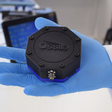
USB Spectrometer

UV-VIS Spectroscopy and Visible Spectroscopy
UV-VIS spectroscopy studies the absorption of ultraviolet and visible light by materials. In molecules and atoms, this absorption corresponds to electronic transitions, and therefore this technique can be used to probe the electronic levels in materials. UV-vis spectroscopy can also cover the NIR region of the electromagnetic spectrum, with UV-vis spectrometers often spanning a spectral range of 190-1,100 nm.
Generally, the property that is actually measured in UV-vis spectroscopy is the transmission of light through a material or the reflectance by it. The sample is illuminated by a light source - this can either be monochromatic or broadband depending on what information is required (see Light Sources for Spectroscopy). The light transmitted through the sample, T, and the light reflected by it, R, is then collected by a detector and compared to a reference. The absorption spectrum, A, can then be calculated through:
Often, either T or R can be assumed to be zero. For example, this would be the case if it is known that the material is mounted onto a substrate with zero transmission in the spectral region of interest. Here, only one property would need to be measured.
A schematic of a typical UV-vis transmission setup is shown in the figure below. This set up shows an optical spectrometer, such as the Ossila USB Spectrometer used in combination with various other optical components. Here, the sample is in solution and held inside a cuvette. An empty cuvette, or a cuvette filled with the sample dissolved in solvent could be used as a reference in this type of measurement. If the sample were a thin film on a substrate, a blank substrate could be used as the reference.
Alternatively, users can take absorbance and transmission measurements using spectrophotometers or spectrofluorometers. These dedicated pieces of equipment contain all the components needed to measure absorbance and transmission in one system.
As well as the energy levels, UV-vis spectroscopy can also give information on the thickness or concentration of a sample through the Beer-Lambert law. The transmission, T, of the sample that has traversed through a material of thickness d is given by:
where I0 and I are the intensity of the light before and after traversing the material, respectively, and α(λ) is the absorption coefficient of the material. This quantity is material-specific and wavelength-dependent (λ). It is related to the wavelength-dependent absorption cross-section, σ(λ), which gives the probability that a photon with wavelength λ will be absorbed by the material and usually has units of cm2, through:
where N is the number of absorbing molecules per unit volume. If the absorption cross-section of the material is known, its concentration can be calculated.
Fluorescence Spectroscopy
Where UV-vis studies the absorption of photons by a material, fluorescence spectroscopy relies on the opposite process - it studies the emission of photons by a material. This will also give information on the possible electronic transitions of the material.
Usually, the sample is excited by a monochromatic source; for example, a continuous-wave (CW) laser diode. The wavelength of the excitation source is usually in the UV/blue region of the EM spectrum to ensure that the excitation photons are of a higher energy than those that will be emitted. The emission from the sample is then detected by a spectrometer, which outputs the data as a function of wavelength. Often, a filter is used to block the excitation light from reaching the detector. This can be an optical filter - for example, a long-pass filter - or a spatial filter.
You can also use spectrofluorometers to accurately conduct fluorescence spectroscopy measurements. These units contain multiple monochromators to isolate light of different wavelengths. This allows users maximum control over both excitation and emission wavelength.
It is important that the sample has a non-zero absorption at the excitation wavelength, otherwise no emission will occur.
Raman Spectroscopy
Raman spectroscopy is generally used to determine the vibrational transitions of a molecule and is often utilized by chemists and pharmacists as a method of identification. It relies on the inelastic scattering of photons by molecules, which results in the molecules gaining vibrational energy and the photons losing it.
This energy (wavelength) shift of the photons is known as the Raman shift. Generally, an infrared laser will be used as the photon source while the Raman spectrum will show intensity (which is proportional to the number of molecules) against shift. This will be given in cm-1 with respect to the laser wavelength. From this, it is possible to determine the structure, composition, and concentration of the sample in question.
Learn More
 Photoluminescent Spectroscopy
Photoluminescence refers to a form of luminescence that results from photoexcitation. Simply, photoluminescence occurs when a material emits light after absorbing a photon from an external light source and is measured using a spectrometer such as an optical spectrometer.
Read more... Fluorescence Spectroscopy
Fluorescence spectroscopy is used to measure fluorescence. The technique often used together with absorbance spectroscopy. Fluorescence is a type of photoluminescence where light is quickly reemitted from a material after incident photons are absorbed. This is different to phosphorescence where there is a delay between photon absorption and emission. The term fluorescence is often used interchangeably with photoluminescence.
Read more...